Positron emission tomography (PET) scan and CT scan

Key points about positron emission tomography (PET) scan and CT scan

  • A positron emission tomography (PET) combined with computed tomography (CT) scan is a 2 in 1 hybrid scan.
  • It's done to provide metabolic or disease specific information about a body organ or system. 
  • It's mainly done to assess cancers, brain diseases and heart-related diseases.
Woman lying on bench about to have PET scan
Print this page

A PET scan uses a special type of camera that detects radiation and a small amount of a radioactive substance (called a radiotracer), to pick up abnormal function or disease in your tissues and organs. It's usually used to examine your entire body from skull to thighs, although some PET scans can be limited to certain organs such as brain or heart only. Different types of radiotracers are used depending on the reason for the scan.  

During the test, the radiotracer is injected into a vein in your arm. As it travels around your body it accumulates in areas where there is a higher level of metabolic activity. If a disease specific radiotracer has been used, it will accumulate in parts of the body where there is a high concentration of the disease being examined. The radiation released by the radiotracer is detected by a PET camera and a highly sophisticated computer reconstruction then produces images of the area being examined.

Images from the PET scanner are generally combined with those of a CT scanner (PET/CT) used at the same time during the procedure. The combined images enable more accurate detection and location of disease.

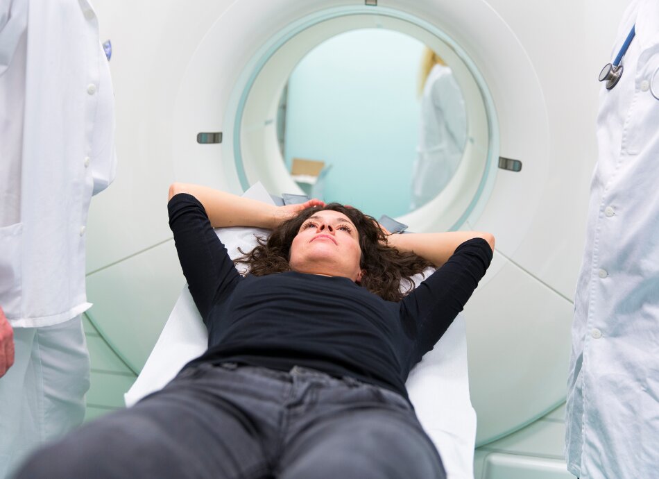
A PET/CT scanner looks like a short tunnel which contains both the PET camera and the CT scan detectors. You will slowly move through the tunnel over a period of 15–30 minutes while the PET camera detects the radiation from your body. A PET/CT scan is carried out by a trained nuclear medicine technologist working with a radiologist or nuclear medicine specialist who specialises in interpretation of PET/CT images and generates a report for your specialist.

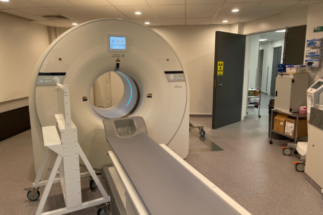
Image credit: Dr Remy Lim

A PET scan is done to help diagnose, stage or monitor a wide range of diseases. These include:

  • Cancer staging or restaging evaluating extent of cancer spread, checking treatment is working or finding a recurrence.
  • Cancer detection – in many different organs, eg, brain, breast, lung, prostate, skin, colorectal, lymph system.
  • Heart disease – finding areas of abnormality, eg, sarcoidosis.
  • Brain disorders – evaluation of brain tumours, Alzheimer's disease and seizures.

Tell your healthcare provider if:

  • you've ever had a bad allergic reaction to a CT contrast fluid
  • you've been sick recently
  • you have a medical condition, eg, diabetes, kidney disease or asthma
  • you’re taking any medicines, herbal supplements or vitamins
  • you’re pregnant or might be
  • you’re breastfeeding
  • you get claustrophobic (scared of small spaces).

You will be given information about what to do to get ready for your PET scan. Usually you will be asked to: 

  • avoid strenuous exercise for a couple of days before
  • stop eating 4 hours before your scan. 

The procedure and the amount of time it takes is different depending on the reason for the scan, and the part of the body being scanned. You’ll be given information about what to expect before you have your scan.

Generally, you will be given the radiotracer injection 45–60 minutes (called uptake time) before the scan and will have to rest quietly while the tracer circulates within your body.

After the appropriate amount of uptake time, you will lie on a padded table which slides into the short tunnel of the PET/CT scanner. It's important that you keep very still so the images are not blurry. The scanner is relatively quiet most of the time. You won’t feel any pain during the scan. Contrast for the CT scan may give you a momentary warm sensation or metallic taste in your mouth.  

The entire procedure usually takes up to 2 hours.

  • The amount of radioactive tracer used during the PET scan is small and will be flushed out of your system in a few hours.
  • Drinking plenty of fluids will help to flush the tracer out of your body.
  • You may be advised to minimise contact with children and pregnant women until the following day.
  • You can get back to your usual activities and diet straight after the scan.

A PET/CT scan is a safe procedure that is regularly performed all around the world. There are no known serious complications of the scan.

The amount of tracer used is small and your body can flush it out of your system in a few hours. During this time it's recommended that you minimise contact with others, especially small children and pregnant women.

You may need to have an injection of a contrast material for the CT scan accompanying your PET scan. As a few people react to the contrast injected, you will be asked about any previous experiences you have had with iodine contrasts before you have your scan. Trained professionals, equipment and medicines are always available in case you have a reaction. Read more about contrast material(external link) and CT scans. 

Contrast material – patient information(external link) RadiologyInfo, US
CT scan(external link) Mayo Clinic, US

References

PET/CT(external link) Pacific Radiology, NZ
PET-CT(external link) Mercy Radiology, NZ
PET scan(external link) Better Health Channel, Australia
Positron emission tomography scan(external link) Mayo Clinic, US, 2021

Need help now?

Healthline logo in supporters block

Need to talk logo

Healthpoint logo

Credits: Healthify editorial team. Healthify is brought to you by Health Navigator Charitable Trust.

Reviewed by: Dr Remy Lim, MBChB, FRANZCR, Medical Director and PET/CT Clinical Lead, Mercy Radiology, Auckland

Last reviewed:

Page last updated: