Echocardiogram

Also called 'echo'

Key points about echocardiogram

  • An echocardiogram, or 'echo' for short, is a type of ultrasound test that's used to examine the structure and functioning of your heart (manawa).
  • The ultrasound waves are turned into moving pictures of your heart that can be seen on a screen and are recorded so your doctor can watch.
  • There are a few different types of echo: 
    • The most common type is the transthoracic echocardiogram (also called a standard echocardiogram).
    • More specialised types of echo are exercise stress echocardiogram, dobutamine stress echocardiogram and transoesophageal echocardiogram.     
Woman sitting in hospital corridor talks to healthcare professional
Print this page

An echocardiogram (echo) is a type of ultrasound (oro ikeike) test that uses high frequency sound waves to examine the structure and functioning of the heart. 

  • The ultrasound waves are turned into moving pictures of your heart that can be seen on a screen and are recorded so your doctor can watch.
  • An echo gives your doctor information about how well your heart pumps and whether your heart valves are working properly. 

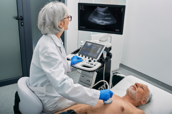
Man having echocardiogram

Image credit: Canva

An echo can be carried out for many different reasons, including to:

  • check how well your heart is working after a heart attack
  • look at how well the valves are moving inside your heart
  • see how well an artificial heart valve is working
  • check if any fluid has collected around your heart
  • check the thickness and movement of your heart wall
  • measure the size and shape of your heart's chambers
  • look for blood clots and tumours inside your heart
  • identify the specific cause of heart failure.

Usually, no special preparations are necessary for a transthoracic or standard ECHO.

  • You can eat or drink as you normally would before the test.
  • Take your medicines as you normally would, they won't affect the test. 
  • You'll need to undress to the waist in a private space and a hospital gown will be provided.

For specialised echocardiograms, there may be specific things you need to do to prepare for the test. You doctor will provide you with more detailed information.

This video provides information about echocardiograms. It may take a few moments to load. 

(British Heart Foundation, UK, 2014)

  • An echo is usually done by a trained technician, in an echo lab.
  • During the echo test, the technician will place a small amount of a cool gel on your chest and a small probe or wand (like a very thick, blunt pen) will be placed on the gel, and moved backwards and forwards over the surface of your chest.   
  • The probe sends sound waves to your heart that then bounce off your heart and make pictures on the screen.
  • If the technician turns on the sound, you may hear a whooshing noise as your heart beats.
  • The technician may ask you to breathe in and out slowly, or to briefly hold your breath during some parts of the test. You may have to turn on to your side during the test so that the technician can scan your heart from different angles.
  • The test will take about 45 minutes.

Sometimes the transthoracic or standard echo doesn't provide enough information and your doctor may request a more specialised echo test.

Exercise stress echocardiogram

During this type of echo you'll be asked to walk on a treadmill or ride an exercise bike while pictures are taken of your heart. This allows your doctor to understand how your heart copes when it's made to work harder, and is helpful to diagnose whether you have angina or not. It can also give your doctor information about the severity of a heart-valve problem.

Dobutamine stress echocardiogram

This type of echo also allows your doctor to understand how your heart copes when it's made to work harder. If you can't exercise, you may be given medicine called dobutamine to make your heart react as if you were exercising. A drip will be put in a vein in your arm, and dobutamine will be infused into the drip, causing your heart to work harder. While this is happening, the technician will take pictures of your heart using an ultrasound probe gently placed on your chest.

Sometimes an even more specialised echocardiogram might be needed, see the section below.
A transoesophageal echo is a more specialised type of echo. With this type, pictures of your heart are taken by inserting a probe through your mouth and down into your throat (oesophagus). These pictures are clearer to see than those produced using a probe on your chest (as in a standard echo) because your oesophagus is close to your heart and there's no chest wall in the way.

Preparation

You'll be given instructions about how to prepare for this type of echo. You shouldn't eat or drink for 4 hours before the test. Stop taking any blood thinning medicines beforehand too – you'll be told when to stop and how long for. You'll need to have a blood test to check your kidney function a few days before the test.

When it's time for the echo, an IV line will be inserted into your arm. This is used to give you a calming medicine as well as saline or a dye to help get a better image. You'll be given an anaesthetic spray to numb your throat and then a thin tube with a probe will be passed down until it's in the right position to record the pictures needed. The tube is removed at the end of the test.

The following links provide further information on echocardiograms. Be aware that websites from other countries may contain information that differs from New Zealand recommendations.

Heart tests(external link)  Heart Foundation NZ
Echocardiogram(external link) Patient Info, UK
Echocardiogram(external link) Mayo Clinic, US

Brochures

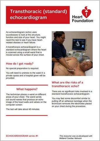
Echocardiogram (echo)(external link) Heart Foundation and Midland Cardiac Network, NZ
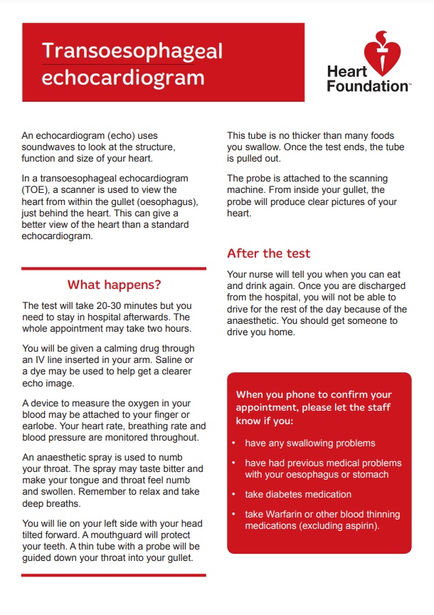
Echocardiogram – transoesophageal(external link) Heart Foundation and Midland Cardiac Network, NZ
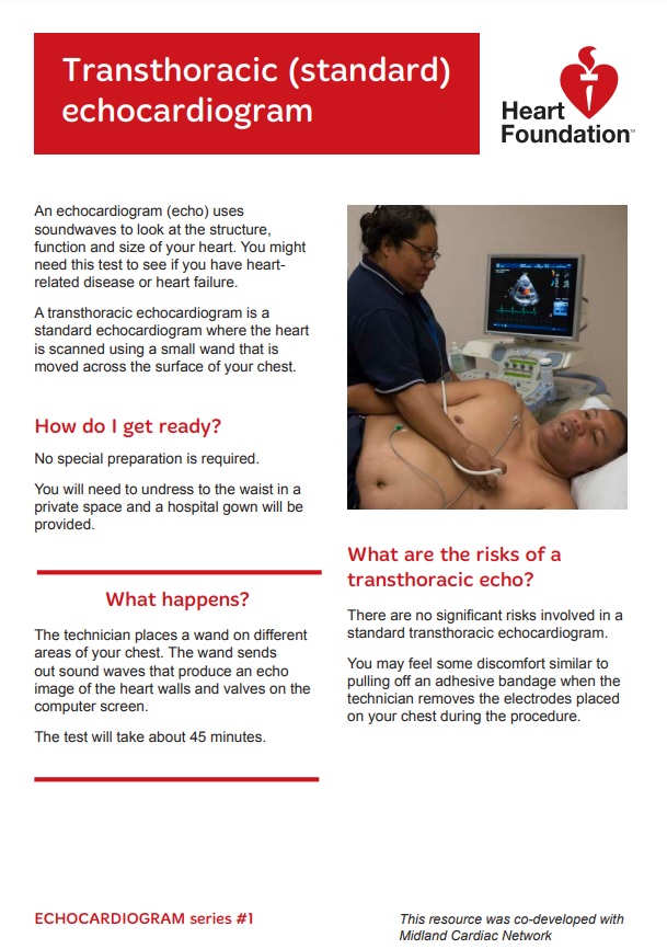
Echocardiogram – transthoracic(external link) Heart Foundation and Midland Cardiac Network, NZ
Dobutamine stress echocardiogram(external link) Heart Foundation, NZ

References

  1. Echocardiogram(external link) Mayo Clinic, US, 2023
  2. Heart tests(external link) Heart Foundation, NZ
  3. Echocardiogram(external link) Patient Info, UK

Brochures

echo

Echocardiogram (echo)

Heart Foundation and Midland Cardiac Network, NZ

echocardiogram transoesophageal

Echocardiogram – transoesophageal(external link)

Heart Foundation and Midland Cardiac Network, NZ

echocardiogram transthoracic

Echocardiogram – transthoracic(external link)

Heart Foundation and Midland Cardiac Network, NZ

Need help now?

Healthline logo in supporters block

Need to talk logo

Healthpoint logo

Credits: Healthify editorial team. Healthify is brought to you by Health Navigator Charitable Trust.

Reviewed by: Claire Salter, Pharmacist, Tauranga

Last reviewed: