(Cancer Research UK, 2015)
Low or no data? Visit zero.govt.nz, scroll down the page then click on our logo to return to our site and browse for free.
Bone scan
Key points about bone scans
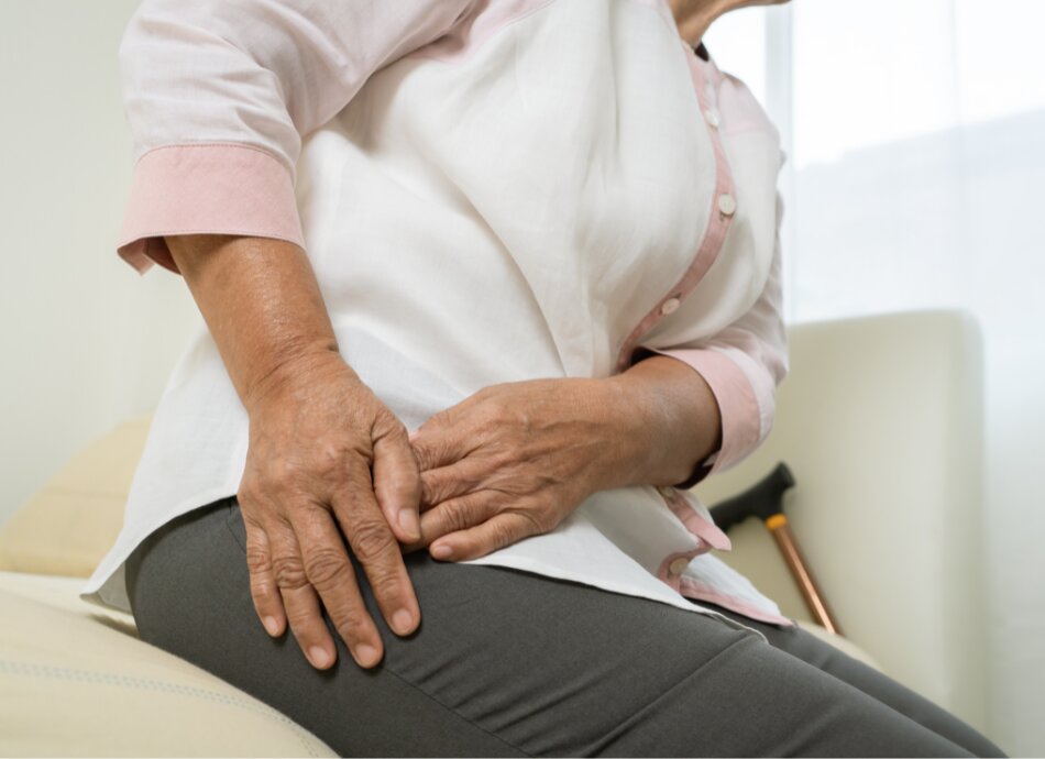
- A bone scan is a test to check for problems inside your bones.
- Your doctor may order a bone scan for you if you have bone pain or they think you might have a problem with your bones, eg, some types of fracture, bone infection, Paget's disease of the bone, bone cancer, unexplained pain or arthritis.
- Bone scans can often find problems days to months earlier than an X-ray can.
When you have a bone scan a tiny amount of radioactive chemical called a radionuclide (also called a radioisotope) is injected into a vein. Your body concentrates this in any parts of your bone that are changing or being repaired. The radionuclide sends out gamma rays which are detected by a scanner.
A bone scan is sometimes called a radionuclide scan, bone scintigraphy or a nuclear medicine bone scan – they're all the same thing. A bone scan is different to a bone density scan (also called a DEXA scan) which is usually used to look for osteoporosis.
When is a bone scan performed?
Your doctor may order a bone scan if you have bone pain and/or if they think you have a problem with your bones, such as:
- Some fractures that are difficult to see on X-ray.
- Bone infection (osteomyelitis) and infections around joint replacements.
- Paget’s disease of the bone.
- Cancers such as bone cancer.
- To see if cancer from other parts of the body have spread to the bones (metastasis).
- Unexplained bone pain.
- Arthritis.
If you're pregnant you can't have a bone scan. If you're breastfeeding you will need to express and discard your milk – the staff doing the scan will let you know what to do.
A bone scan doesn't need any special preparation.
- Wear loose, comfortable clothing. Avoid clothes that have zips, belts or buttons made of metal.
- You can eat and drink normally.
- Take your usual medication unless you're told otherwise.
- Expect the visit to take about 6 hours.
A bone scan is done in a hospital, in the nuclear medicine, medical physics or radiology department. The scan is normally done by a nuclear medicine technologist or radiographer (a technician trained to do X-rays and other scans).
Before your scan you'll be asked to remove jewellery, removable dentures or plates, glasses and any metal objects or clothing that might interfere with the scanner. You may be given a gown to wear.
The radiographer will explain what's going to happen and will ask you to sign a consent form. They will then inject a small amount of a radionuclide into a vein in your arm. The radionuclide travels through your bloodstream and into your bones. It takes about 2 to 3 hours to be absorbed into your bones. You may be allowed to leave the hospital during this time.
When it's time to do your scan, you'll be asked to lie on a table where a large camera called a scanner will measure how much of the radionuclide has been absorbed by your bones. You will be asked to move into different positions and hold very still so the camera can take pictures of all sides of your body. You may need to lie still for as long as 30 minutes. The radionuclide collects more in parts of the bone that are breaking down and repairing themselves, these show up darker on the scan and are called hot spots. A digital picture is made by the scanner which a radiologist or specialist uses to work out what's happening.
A bone scan itself takes about 1 hour.
The amount of radionuclide that's absorbed depends on the amount of growth or activity in the bone.
-
-
- Test results are normal when the radionuclide is spread evenly throughout your body. This means that you're unlikely to have any major bone problems.
- Areas that absorb little or no amount of radionuclide appear as dark or "cold" spots. This could show a lack of blood supply to your bone, or certain types of cancer.
- Areas of bone growth absorb more radionuclide and show up as "hot" spots in the pictures. These may mean you have a problem (eg, cancer, a fracture or an infection).
-
You won't get your results on the day of the scan. The doctor who ordered your scan should discuss your results with you at the next appointment.
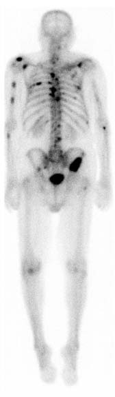 Image credit: RadsWiki(external link) via Wikimedia Commons
Bone scans don't generally cause any after effects. The radionuclide loses radioactivity over time. It passes out of your body through your urine over about 24 hours. You will need to drink plenty of water for a day after the scan to help flush the radionuclide out of your body. You will need to take special care after passing urine, like flushing the toilet twice and washing your hands thoroughly.
A radioactive injection sounds dangerous, but only involves a tiny amount of radiation. For many scans it's about the same amount as having 2 normal X-rays. You can talk to the doctor who is ordering your bone scan if you're worried about this, or with the radiographer before your scan. As with all chemicals it's possible to be allergic to the radionuclide, and as with all injections bruising can happen.
Bone scan(external link) Patient Info, UK
Bone scan(external link) Cancer Research, UK
Nuclear medicine bone scan(external link) Inside Radiology, Australia
Bone scan(external link) Mayo Clinic, US
References
- Bone scan(external link) Patient Info, UK
- Bone scan(external link) Cancer Research, UK
- Nuclear medicine bone scan(external link) Inside Radiology, Australia
- Nuclear medicine(external link) Inside Radiology, Australia
- Bone scan(external link) Mayo Clinic, US, 2022
Need help now?

Credits: Healthify editorial team. Healthify is brought to you by Health Navigator Charitable Trust.
Reviewed by: Dr Emma Dunning, Clinical Editor and Advisor
Last reviewed:
Page last updated: